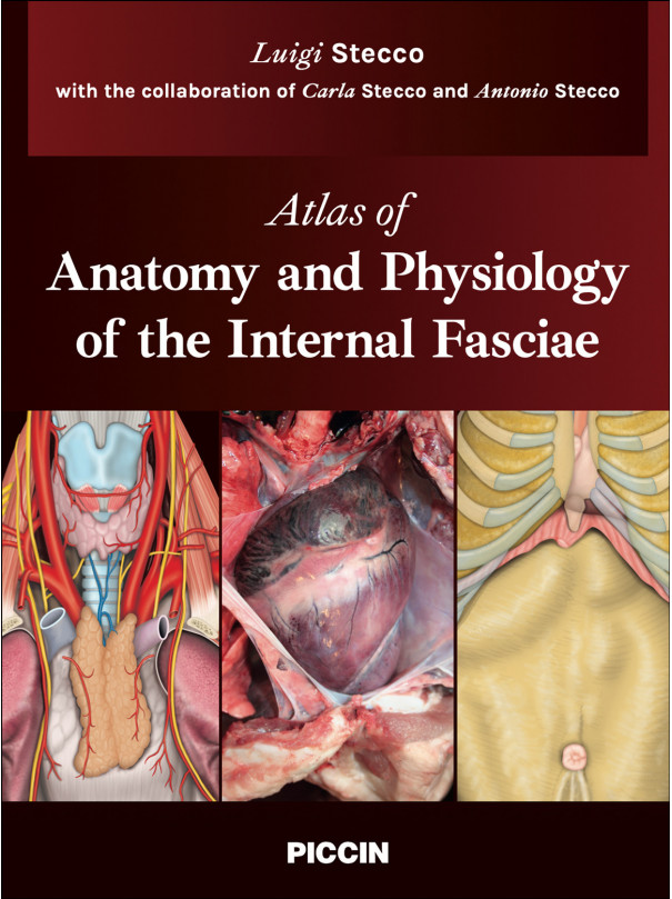
Atlas of Anatomy and Physiology of the Internal Fasciae
Luigi Stecco with the collaboration of Carla Stecco and Antonio Stecco
88,00 €
FOREWORD
After 20 years of clinical practice experience, I got acquainted with the method “Fascial Manipulation” by Luigi Stecco and was amazed by the possibilities that the method opens up for the treatment, rehabilitation and prevention of injuries of the musculoskeletal system and dysfunctions of internal organs.
The method “Fascial Manipulation” is based on fundamental knowledge in the field of human anatomy and physiology and it is very encouraging that scientific research continues today, enriching clinical experience.
The presented knowledge and results force us to completely rethink the anatomy and functions of the autonomic nervous system. The role of internal fasciae in the correction of functional disorders of internal organs prove that the sympathetic and parasympathetic nervous systems are not antagonists.
During the rehabilitation period after musculoskeletal surgery, as a rule, the emphasis is made on physiotherapy and kinesiotherapy. However, this does not always allow to achieve a long-lasting result – patients often start to complain of various internal organs dysfunctions.
This problem can be solved by understanding and consequent clinical application of the internal biomechanical model presented by Luigi Stecco, which is based on the motility and mobility of internal organs directly related to the autonomic nervous system and, of course, to the state of the fascial tissue of internal organs and muscles. This understanding makes the use of peristaltic therapy very effective. Proper coordination of the autonomic nervous system, three types of nerves – splanchnic nerve, vagus nerve, phrenic nerve – and fascial tissues improves peristalsis of internal organs by increasing their living space.
The brain must have an accurate and effective perception of the body which is very important for its survival, therefore, thanks to reflexology, manual therapy of the superficial fascia improves the vascularization of the dermatomere, adipotomere and lymphatomere and, as a result, there is an improvement of afferent and efferent interactions of surface structures with the central nervous system.
Despite the scientific depth, Luigi Stecco’s proposed concept is characterized by simplicity and logic. This knowledge will be useful for specialists from different medical fields and will allow achieving long-lasting results in the treatment and rehabilitation of patients.
I am sincerely grateful to Luigi Stecco, Antonio Stecco, Carla Stecco and other scientists in this field for their advanced knowledge, valuable practical experience and dedication to their mission - the preservation and restoration of human health.
ALEKSANDR STOGOV, MD
Orthopaedic Doctor,
Certified Fascial Manipulation specialist,
Moscow, Russia
INTRODUCTION
The driving force behind writing this Atlas has been the opportunity to publish new images of the fasciae of human internal organs and to compare them with images of the internal fasciae of other animals. We are also excited to present new concepts regarding the physiology of organ-fascial units, apparatus-fascial sequences, systems and, in particular, of the autonomic nervous system.
This Atlas is divided into three parts and consists of a total of twelve chapters.
The first part starts from the first chapter, which presents the internal fasciae and a new interpretation of the microscopic autonomic nervous system. The second, third, fourth and fifth chapters address the organs within the neck, thorax, lumbar and pelvic segments. The organs within each one of these segments are united together by fasciae to form the glandular, visceral and vascular organ-fascial units (o-f unit).
The sixth chapter introduces the second part, which is dedicated to the apparatus-fascial sequences and to the autonomic nerves that originate from the central nervous system (CNS). The seventh, eighth, ninth and tenth chapters address the fasciae that connect the glandular, visceral, vascular and receptor o-f units together in a longitudinal direction.
The third part starts from the eleventh chapter, which is dedicated to the superficial fascia and the macroscopic autonomic nervous system formed by the paravertebral and prevertebral ganglia.
The twelfth chapter addresses the development and the functions of the three external systems (cutaneous, adipose and lymphatic) and the three internal systems (thermoregulatory, metabolic and immune).
Our literature research regarding the autonomic nervous system (ANS) encountered some difficulties because different texts use different terms to describe the same structure. For example, in Chiarugi’s anatomical text (1975), the first lines of the chapter regarding the ANS state: “Due to its anatomical connections, the autonomic or visceral or vegetative or sympathetic or greater sympathetic nervous system should be considered as an offshoot of the cerebrospinal system. The autonomic system includes I) the thoracolumbar or sympathetic system, II) the craniosacral or parasympathetic system and III) the intramural or metasympathetic system.” On the contrary, as in many other anatomical texts, the physiology text by Saladin (2019) sustains that the ANS consists of two subsystems:
the sympathetic system and the parasympathetic system. In English language texts, the term “sympathetic” is synonymous with the term “orthosympathetic”, whereas Chiarugi uses the term “sympathetic” as synonymous to the “autonomic system”. In this Atlas, the terms “sympathetic” and “autonomic nervous system” (ANS) will be used to indicate the entire autonomic system.
According to which prefix (ortho, meta, adeno or para) is associated with the suffix “sympathetic”, the meaning is modified as follows:
■ metasympathetic system: indicates the intramural neuronal network that has the role of activating smooth muscles within the walls of the internal organs (myenteric autonomic nervous system)
■ orthosympathetic system: excitomotor stimuli that are conducted by splanchnic nerves and are directed to the kidneys and the smooth muscles of the blood and lymphatic vessels
■ adenosympathetic system: excitomotor stimuli that are conducted, in part, by the phrenic nerve and are directed to the glandular capsules and glands (from Greek, aden = gland)
■ parasympathetic system: excitomotor stimuli conducted by the vagus nerve and, in particular, directed to the intramural ganglia of the respiratory and digestive apparatus.
The autonomic system can also be divided into two parts according to size and function:
■ the microscopic autonomic system, which, in turn, includes the intramural or metasympathetic neuronal network and the extramural ganglia situated in the insertional fasciae
■ the macroscopic autonomic system, which, in turn, includes the paravertebral and prevertebral autonomic ganglia. These ganglia connect with the CNS via the vagus, phrenic and splanchnic nerves.
The macroscopic ganglia are influenced by stimuli coming from the medulla oblongata and the hypothalamus. Hence, they do not have a completely autonomous activity because they act in response to stimuli that come from the CNS.
No customer comments for the moment.


